Staying Vigilant with Mass Aspirates
Should you really get EVERY lump checked? Dr. Susan Ettinger, DVM, Dip. ACVIM (Oncology) explains why we should all stay vigilant with mass aspirates and get everything checked.

Read Time: 5 minutes
I am a massive advocate of aspirating every lump and bump on your dog, even though many turn out to be benign cysts or lipomas (fatty tumors). The story below will illustrate why I’m so vigilant, but first, a little about mass aspirates.
Mass Aspirate: What Is It?
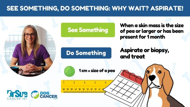
When there’s a lump or bump on the surface or just under your dog’s skin, and you can feel it, you should get it aspirated. It’s a simple test, and no anesthesia is required. A very thin needle is put into the lump, and then whatever fluid is inside is drawn up into the syringe, and your vet or a lab can take a look and see what’s in it. This is a really simple screening test for many types of cancer. If something problematic turns up, you can move quickly to address it.
On the other hand, if the masses are benign and not bothering your dog, they probably do not need to be removed: I typically recommend leaving them alone and monitoring them in the future. Perhaps I will recommend surgically removing them at the same time as another procedure like dentistry, but I will rarely recommend removal if the aspirate was benign, because going under anesthesia has its own risks, too.
Your primary care vet and you are on the front line of skin and subcutaneous mass monitoring. Dogs usually come to me, the oncologist, after the aspirate or biopsy has been performed. They either already have a diagnosis of cancer, or at least suspicious for malignancy.
Why Fine Needle Aspirates Matter
Here’s the story I promised, and why I’m so vigilant about aspirates for my own patients.
One of the ways I give appreciation to my wonderful oncology nurses is by monitoring the health of their pets. It’s my pleasure to care for them, including aspirating any lump or bump.
One of my nurses has an amazing white Pitty (Pit Bull), named Smokey, which I adore. I have aspirated numerous skin masses on numerous occasions over the years. And until two weeks ago, the masses have always been fatty deposits.
When my nurse mentioned she was bringing Smokey in to check out a new mass earlier this month, we all expected it to be the routine: quick aspirate, fat on the microscope slide, document it in is his records, give a treat to Smokey, collect some wags and kisses, and on his way. Actually the day she 1st brought him in was an insane clinic day that kept us all on the move, shorter lunch breaks, overtime for the nurses … and I missed storytime with my two boys. We never did get to Smokey. No worries, my nurse said. She would bring him in on Monday.
On Monday I did my exam. Smokey had a 5 cm mass that was deeply attached to the underlying tissue on his left flank area. As I did my aspirate, I could see blood collecting in my needle and syringe. I immediately knew this was not a lipoma. I aspirated the mass in a few more areas, and we submitted the slides to the lab for cytology.
I asked, but there was no history of trauma to explain a mass with blood inside on his trunk. I told my nurse my clinical hunch was a blood vessel tumor: either a benign hemangioma or the malignant version, hemangiosarcoma. Tears welled up in her eyes, and Smokey became concerned that his mom was upset. My nurse deals with cancer in dogs and cats every day, but it is so different when it is YOUR dog. I could see her mind start to race and shut down at the same time. I gave her a huge hug, and we waited anxiously overnight for his cytology results.
The Aspirate Found … Soft Tissue Sarcoma
The aspirate came back: it was a soft tissue sarcoma. I reminded her about soft tissue sarcomas, or STS. They develop in a variety of connective tissues, muscles, and fat. They can be found in sites all over the body, from head to trunk to paws. The majority of these tumors are usually aggressive locally, which means they invade the neighboring tissues. They are also prone to recur if they are not removed with wide margins. The good news is that the low and intermediate-grade tumors typically don’t metastasize, or spread.
I know it’s weird to hear me say this, but if you had to pick a malignant tumor for your dog, this is a pretty good choice. Having soft tissue sarcomas is not the worst-case scenario for your dog. Low and intermediate grades of soft tissue sarcomas are very treatable and have excellent long-term survival rates (some up to five years). The recommended treatments include surgery and/or radiation for low and intermediate grades. And, occasionally, chemotherapy.
Dealing with the Tumor
The next step was a PRE-surgical biopsy to confirm the tumor type and help our soft tissue surgeon appropriately plan the surgery. A biopsy confirmed a low-grade hemangiopericytoma, a type of STS. We had already run blood and urine tests, and we checked for metastasis with chest X-rays and abdominal ultrasound – all clear!
On surgery day, Smokey had a CT scan to determine the extent of the tumor better than we could feel. These tumors are infamous for having tentacle-like projections that can extend for centimeters, far from the main mass. If we aren’t careful and leave the tumor tentacles in his tissues, we would have incomplete margins, which would mean his tumor would be ten times more likely to recur.
This is why imaging before surgery, as close to surgery as possible, is so important.
The surgeon reviewed the CT scan as Smokey was prepped for surgery. She reported the mass was most likely resectable (she could remove it), but she would also have to move a flap of skin to close the incision once she was done. You see, because of those tentacles, these tumors require 3 centimeter (more than an inch) margins around the tumor. Not just the skin, either — 3 centimeters deep into the body, too. That is a really big surgery: for a 5-centimeter tumor, the resulting scar should be at least 11 cm (or 4.3 inches).
Happy Outcome
Smokey’s surgery was a big one, but it went well. He spent 2 days in our ICU recovering, and then we waited for the post-surgical biopsy report to come back and tell us how we did.
Well, I got the biopsy report back this weekend. GREAT NEWS: a low grade (grade 1) hemangiopericytoma with wide, clean margins. This is great news because no post-operative radiation of chemotherapy is recommended. We’ll just do regular monitoring of the scar and the rest of his body.
This story illustrates why there is so much information in the Guide about soft tissue sarcomas. Not all stories end this well, and it’s usually because we just don’t realize how important it is to get every lump and bump checked out with an aspirate.
Stay Vigilant About Lumps and Bumps
Just because your dog has had multiple lipomas or other benign masses in the past, don’t get too relaxed. Lumps and bumps are not always benign, as Smokey proved. After dozens of benign aspirates, it only took one to produce a cancer diagnosis. Stay vigilant and have those lumps and bumps aspirated. It’s not a big deal for the dog (think about as bad as a needle) and it is worth knowing what you’re facing.
Smokey had to wait an extra four days for treatment for his cancer because we all got so busy and put his aspirate on the back burner. Do I think the delay was detrimental to his outcome? Ultimately, no — but he’s a lucky dog with an oncology nurse and the resources of one of the best hospitals at his disposal.
So tonight, and every night, play with and pet your dog and take notice of any masses (lumps or bumps) … and get them checked out. No one — not a vet, not an oncologist, and not you — can tell what a lump is just by feeling. And “waiting and seeing” is not a good idea. Get the masses aspirated. Don’t assume it’s another lipoma. The earlier we find tumors, the better.
To your dog’s health,
Dr. Sue
Editorial Note: This post was originally published on a retired blog about dog cancer.
Sue Ettinger, DVM, Dip. ACVIM (Oncology)
Topics
Did You Find This Helpful? Share It with Your Pack!
Use the buttons to share what you learned on social media, download a PDF, print this out, or email it to your veterinarian.


